


Written by Ms Agnes Grzesik
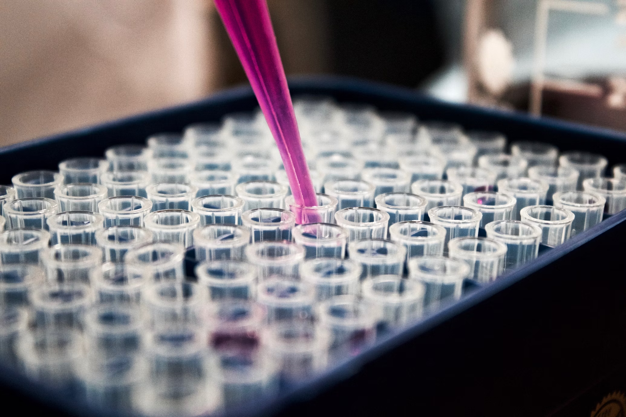
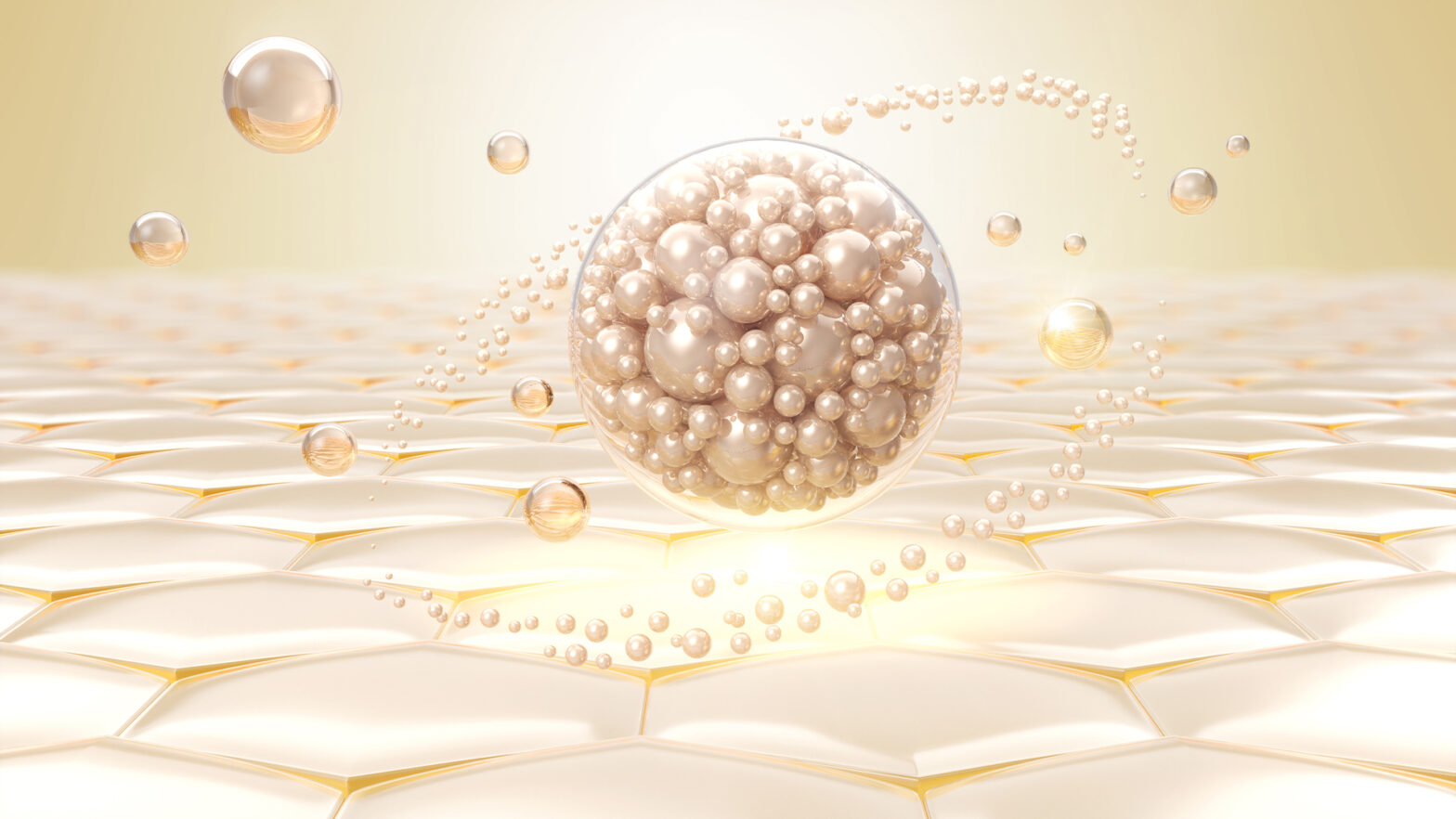
Exosomes are extracellular vesicles described 40 years ago, first noted in ferritin receptor transfer mechanism from studies on the loss of transferrin during the maturation of reticulocytes into erythrocytes (Harding, Heuser, & Stahl, 1983). Since then, exosomes have been implicated in cell-cell communication involving homeostatic mechanisms, which when disrupted may induce disease. Exosomes are released from cells upon fusion of an intermediate endocytic compartment, the multivesicular body (MVB), with the plasma membrane. This liberates intraluminal vesicles (ILVs) into the extracellular milieu as exosomes (Johnstone, Adam, Hammond, Orr, & Turbide, 1987). Their size is typically 40–100 nm and are released from cells for a variety of reasons, which have been overlooked.
Exosomal vesicles are important due to the fact they are capable of transferring intracellular proteins into the cytoplasm, which could affect nearby cells, thus transferring disease signals throughout an organ system. This is depicted in Figure 1, where exosomal bodies exit the infected B lymphocyte carrying intracellular moieties that could affect nearby cells in the lymph nodes.

This form of communication, especially between T and B lymphocytes was deemed important for direct cell-cell communication, and transmission, between cells. Some of the earliest studies in exosomes showed that MHC-antigen complexes, which are important for eliciting an immune response, could be recognised by T lymphocytes, orchestrating an effective antitumor immune response (Raposo et al., 1996). It was shown that exosome-based vaccines may be the key to suppressing tumour growth (Zitvogel et al., 1998). In light of these findings, exosomes can be important in transferring a plethora of molecules such as lipids, proteins and DNA (and mRNA) directly into the cells. As such they are ideal carrying vectors suitable for vaccine formation because are composed of cell membranes and not synthetic polymers, fitting a high-level tolerance from the host.
The biology of exosomes allows for their exploitation in rejuvenation and regeneration studies. The latter aims to restore the original structure and functionality of a diseased organ. This also applies to the accumulated damage that occurs during ageing. As such, exosomes could be ideal carriers of molecules that could instruct old and/or damaged cells to return to their healthy stage. It is now evident that cells, especially progenitor/stem cells are capable of a certain plasticity allowing them to revert to a younger/healthier state when the appropriate information is provided (Teixeira, Carvalho, Sousa, & Salgado, 2013). One potential application for exosomes, with direct results, would be skin repair and/or rejuvenation. The skin is frequently injured by acute and chronic wounds, involving age-related diseases and conditions such as diabetes. In vitro studies of human cells showed that exosomes alone could facilitate wound healing by promoting collagen types I and II synthesis, as well as angiogenesis, two milestones for skin regeneration (J. Zhang et al., 2015). From a mechanistic standpoint, the exosomal transfer of Wnt4 and the activation of the AKT pathway was associated with cutaneous skin healing in a rat skin burn model (B. Zhang et al., 2015).
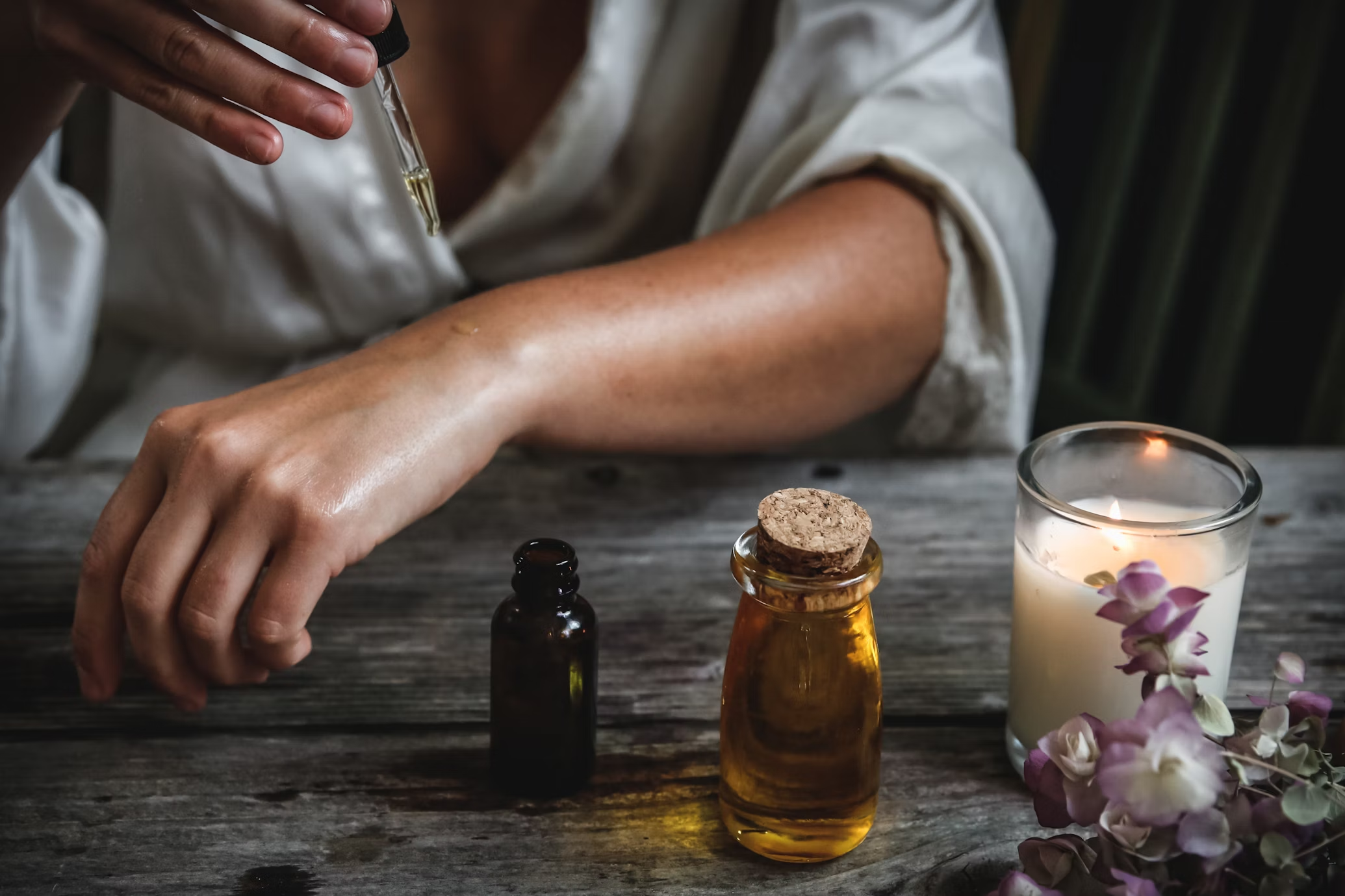
Applications of exosomes could also be used as vectors for delivering molecules that benefit the target-organ in a transient manner. The term cosmeceuticals labels biologically active ingredients that are applied (on the skin) with medical benefits. Currently, liposomes are frequently used as carrying vectors for topical drugs, in the form of lotions, creams, and ointments (Madsen & Andersen, 2010). Liposomes and exosomes share several similarities, yet exosomes may surpass liposomes in delivery efficiency and with significantly reduced side effects (Antimisiaris, Mourtas, & Marazioti, 2018). One major difference is that liposomes are composed of one lipid bilayer, while exosomes have a complicated structure that involves cell membrane proteins, allowing them to interact more naturally with the target cells, i.e. skin cells. This allows exosomes to exhibit a higher specificity while minimising unwanted side effects due to increased immunocompatibility (when derived from an autologous source). In the complex cellular microenvironment of cutaneous skin repair, exosomes have shown promising results, from immune-cell-derived exosomes (Kim et al., 2019; Tienda-Vazquez et al., 2023). In scar remodelling it has been shown that delivery of mRNA molecules through via exosomes could drive white blood cells towards a non-fibrotic skin repair (Chen et al., 2019), while accelerating wound healing and reversing photoaging-related skin appearance (L. Hu et al., 2016; S. Hu et al., 2019).
In conclusion, exosomes are a promising biomedical tool for effectively delivering bioactive molecules in cutaneous skin cells, which may promote their rejuvenation and healing, without serious side effects. Their manufacturing cost is still high, yet gradually decreasing, due to new sources from where they can be extracted.
Contact Details
Ms Agnes Grzesik
Founder & Adult Nurse Practitioner MSN
Preventative, Integrative and Naturopathic Medicine MSN
Booking in bio /or DM on (+44) 07903568573
www.divineskinscience.com
References
Antimisiaris, S. G., Mourtas, S., & Marazioti, A. (2018). Exosomes and Exosome-Inspired Vesicles for Targeted Drug Delivery. Pharmaceutics, 10(4). doi: 10.3390/pharmaceutics10040218
Chen, J., Zhou, R., Liang, Y., Fu, X., Wang, D., & Wang, C. (2019). Blockade of lncRNA-ASLNCS5088-enriched exosome generation in M2 macrophages by GW4869 dampens the effect of M2 macrophages on orchestrating fibroblast activation. FASEB J, 33(11), 12200-12212. doi: 10.1096/fj.201901610
Edgar, J. R. (2016). Q&A: What are exosomes, exactly? BMC Biol, 14, 46. doi: 10.1186/s12915-016-0268-z
Harding, C., Heuser, J., & Stahl, P. (1983). Receptor-mediated endocytosis of transferrin and recycling of the transferrin receptor in rat reticulocytes. J Cell Biol, 97(2), 329-339. doi: 10.1083/jcb.97.2.329
Hu, L., Wang, J., Zhou, X., Xiong, Z., Zhao, J., Yu, R., . . . Chen, L. (2016). Exosomes derived from human adipose mensenchymal stem cells accelerates cutaneous wound healing via optimizing the characteristics of fibroblasts. Scientific Reports, 6, 32993. doi: 10.1038/srep32993
Hu, S., Li, Z., Cores, J., Huang, K., Su, T., Dinh, P. U., & Cheng, K. (2019). Needle-Free Injection of Exosomes Derived from Human Dermal Fibroblast Spheroids Ameliorates Skin Photoaging. Acs Nano, 13(10), 11273-11282. doi: 10.1021/acsnano.9b04384
Johnstone, R. M., Adam, M., Hammond, J. R., Orr, L., & Turbide, C. (1987). Vesicle formation during reticulocyte maturation. Association of plasma membrane activities with released vesicles (exosomes). J Biol Chem, 262(19), 9412-9420.
Kim, H., Wang, S. Y., Kwak, G., Yang, Y., Kwon, I. C., & Kim, S. H. (2019). Exosome-Guided Phenotypic Switch of M1 to M2 Macrophages for Cutaneous Wound Healing. Adv Sci (Weinh), 6(20), 1900513. doi: 10.1002/advs.201900513
Madsen, J. T., & Andersen, K. E. (2010). Microvesicle formulations used in topical drugs and cosmetics affect product efficiency, performance and allergenicity. Dermatitis, 21(5), 243-247.
Raposo, G., Nijman, H. W., Stoorvogel, W., Liejendekker, R., Harding, C. V., Melief, C. J., & Geuze, H. J. (1996). B lymphocytes secrete antigen-presenting vesicles. J Exp Med, 183(3), 1161-1172. doi: 10.1084/jem.183.3.1161
Teixeira, F. G., Carvalho, M. M., Sousa, N., & Salgado, A. J. (2013). Mesenchymal stem cells secretome: a new paradigm for central nervous system regeneration? Cell Mol Life Sci, 70(20), 3871-3882. doi: 10.1007/s00018-013-1290-8
Tienda-Vazquez, M. A., Hanel, J. M., Marquez-Arteaga, E. M., Salgado-Alvarez, A. P., Scheckhuber, C. Q., Alanis-Gomez, J. R., . . . Parra-Saldivar, R. (2023). Exosomes: A Promising Strategy for Repair, Regeneration and Treatment of Skin Disorders. Cells, 12(12). doi: 10.3390/cells12121625
Zhang, B., Wang, M., Gong, A., Zhang, X., Wu, X., Zhu, Y., . . . Xu, W. (2015). HucMSC-Exosome Mediated-Wnt4 Signaling Is Required for Cutaneous Wound Healing. Stem Cells, 33(7), 2158-2168. doi: 10.1002/stem.1771
Zhang, J., Guan, J., Niu, X., Hu, G., Guo, S., Li, Q., . . . Wang, Y. (2015). Exosomes released from human induced pluripotent stem cells-derived MSCs facilitate cutaneous wound healing by promoting collagen synthesis and angiogenesis. J Transl Med, 13, 49. doi: 10.1186/s12967-015-0417-0
Zitvogel, L., Regnault, A., Lozier, A., Wolfers, J., Flament, C., Tenza, D., . . . Amigorena, S. (1998). Eradication of established murine tumors using a novel cell-free vaccine: dendritic cell-derived exosomes. Nat Med, 4(5), 594-600. doi: 10.1038/nm0598-594