


Scientist have discovered tumors in the bones of mummies in ancient Egypt and references to the disease have been found in manuscripts from that time suggesting that cancer has been a part of human existence dating back to around 3000BC. Ancient Egyptian textbooks on trauma surgery describe what looks to be cases of breast cancer and there is reference to the disease being untreatable.
Since then, we have come a long way in the detection and treatment of cancer, with advances like the Pap Smear and mammograms leading to much earlier diagnosis and treatments such as hormone therapy and chemotherapy playing a major role in the treatment of cancer.
What is Flow Cytometry?
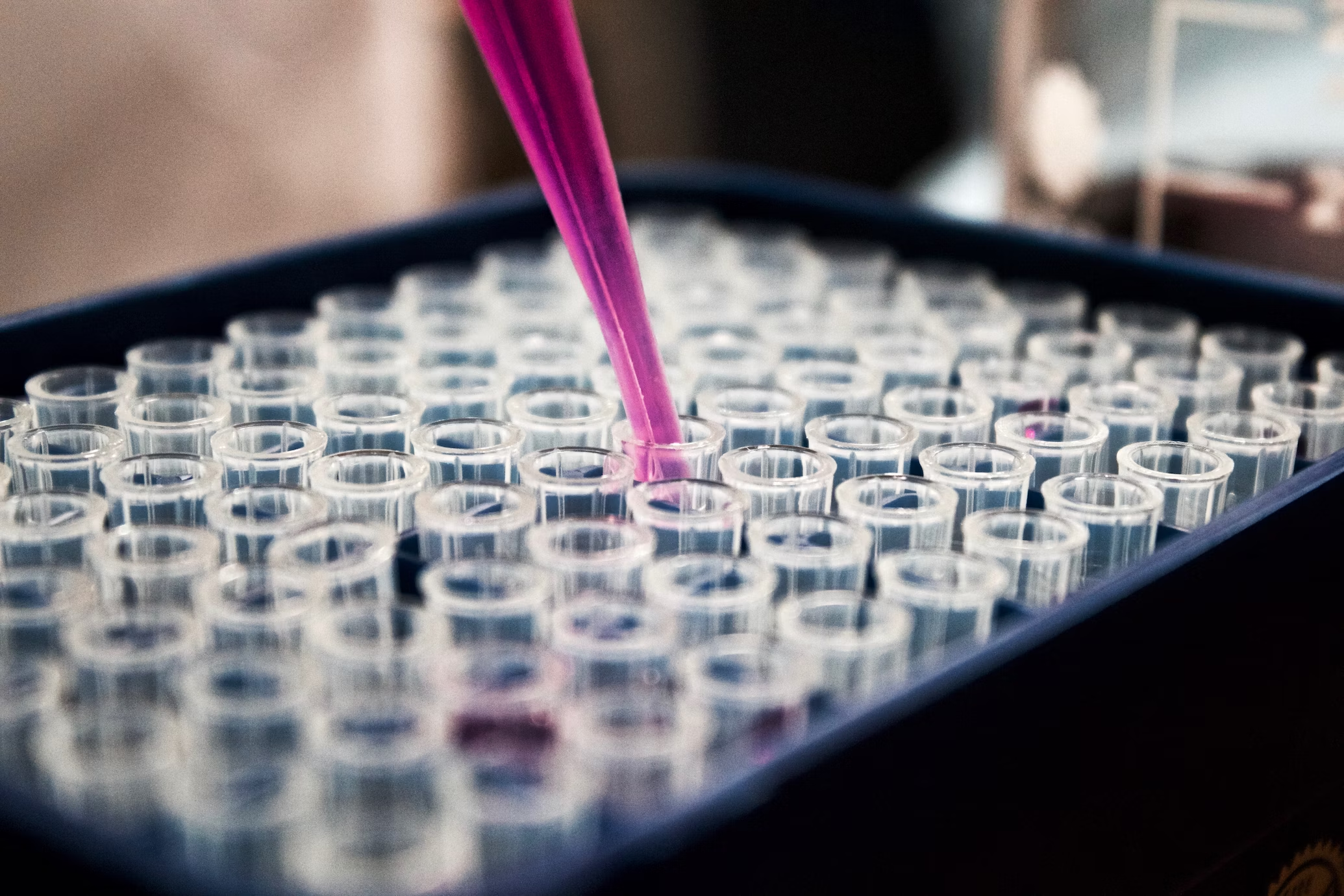
One technological advancement is flow cytometry, an analytical tool which assists medical professionals in the diagnosis, classification and management of cancer. According to the National Cancer Institute, flow cytometry is ‘A method of measuring the number of cells in a sample, the percentage of live cells in a sample, and certain characteristics of cells, such as size, shape, and the presence of tumor markers on the cell surface. The cells are stained with a light-sensitive dye, placed in a fluid, and passed in a stream before a laser or other type of light. The measurements are based on how the light-sensitive dye reacts to the light.’
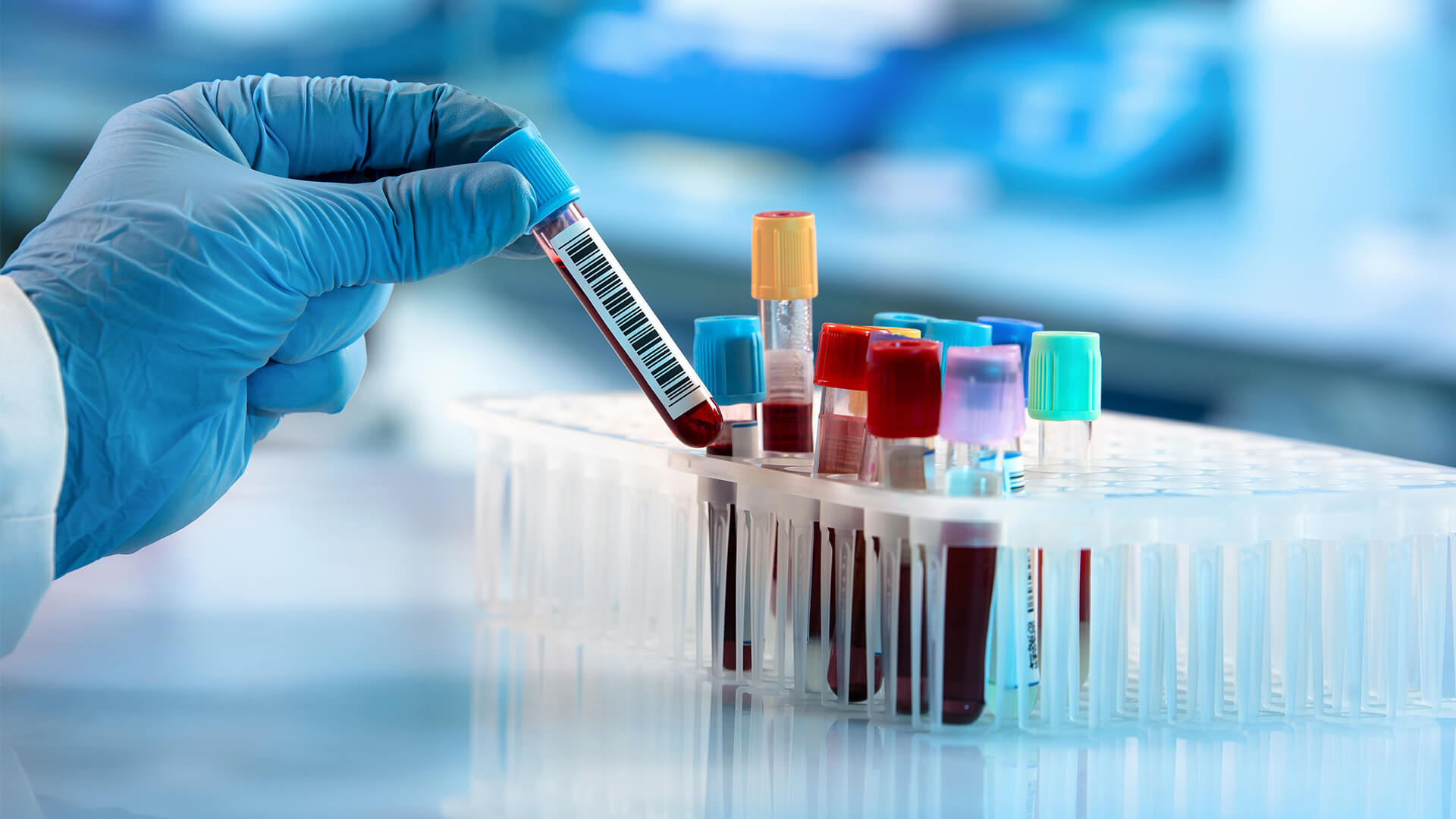
Bone marrow, lymph nodes and blood samples are all examined and tested using a flow cytometry machine. This has proven to be the most accurate way of telling lymphomas from non-cancer diseases in the lymph nodes.
How Does It Work?
A sample of cells from a biopsy, cytology specimen or blood specimen is treated with antibodies. These antibodies will only stick to the cells that have the antigens that fit with it. These cells are then passed through a laser beam and the cells with the new antibodies will give off a light which can then be measured and analyzed by a computer.
What Do the Results Show?
Once the cells have been analyzed, the lab will look at whether the cells in the sample all have the same substance on their surface. This would suggest that they all come from a single abnormal cell which is likely to indicate the presence of cancer. Alternatively, the discovery of several different cell types with a variety of allergens is likely to indicate that there is no cancer present.
As well as this, flow cytometry can also measure the amount of DNA in cancer cells (referred to as ploidy). Cells are treated with a special dye that in turn triggers a reaction with the DNA. If there is a normal amount of DNA, then the cells are said to be diploid but if the amount is abnormal the cells are described as aneuploid. Aneuploid cancers tend to grow and spread faster than diploid ones.
As well as this, flow cytometry can also be used to measure the percentage of cells in a sample that are in a stage of cell division referred to as the S-phase. The cancer is more likely to be growing faster and more aggressively the more cells that are in the S-phase.